TECHNOLOGIES
Our clinic is equipped with cutting-edge technologies, machinery, and services. Innovations in diagnostic and therapeutic fields aim to improve both dental care and patient comfort.
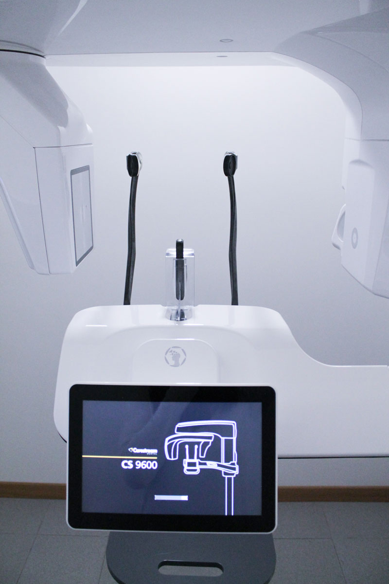
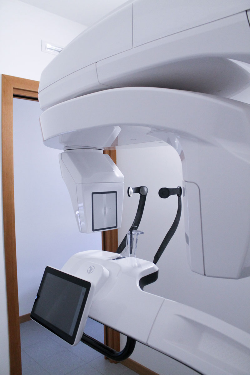
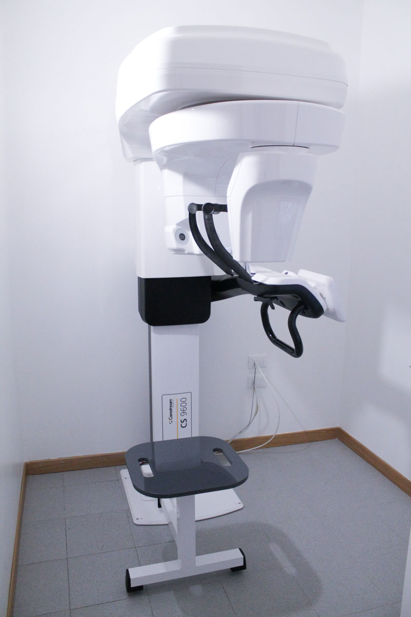
Cone Beam Computed Tomography (CBCT)
The Cone Beam Computed Tomography (CBCT) is a state-of-the-art machine that allows for the reproduction of sections or the generation of a three-dimensional image of the area of interest using radiation acquired by a digital sensor and processed by a computer. This equipment can acquire the entire volume to be examined with a single 360° rotation of the radiogenic tube and detectors, made possible by the emission of a conical-shaped radiation beam and a large-surface panel detector. The images obtained are of high resolution and correspond 1:1 to the anatomical reality.
Advantages of CBCT:
- Short exam duration
- Reduced radiation dose, achieved through the “pulsed” technique that significantly reduces patient exposure time
- Reduced metal artifacts
- Minimal space requirement
The CBCT exam ensures extreme comfort, as it can be performed while sitting or standing, requiring the patient to remain as still as possible for just a few seconds. The exam duration can vary from 10 to 20 seconds depending on the diagnostic information needed. The machine is open, eliminating any claustrophobic issues for the patient.
Intraoral Scanner
Intraoral scanners are powerful devices for capturing optical impressions. Conventional physical impression taking with trays and materials (alginates, silicones, polyethers) can be stressful and uncomfortable for patients, particularly those with a heightened gag reflex. Traditional impression taking can also be challenging for the clinician, especially with technically complex impressions (e.g., for fixed arches on implants). The intraoral scanner, using a light beam (structured light or laser), resolves these issues: it is well tolerated by patients as it does not require conventional materials and is technically simpler for the professional.
The introduction of intraoral scanners in dentistry has completely revolutionized diagnostic and therapeutic protocols, enabling:
- Immediate quality control of the impression
- Elimination of physical storage for plaster models
- Creation of a virtual warehouse
- Elimination of traditional impression materials
- Elimination of the gag reflex issue
- Immediate file transfer to the lab
- Reduced waiting times
- Fewer appointments for the patient
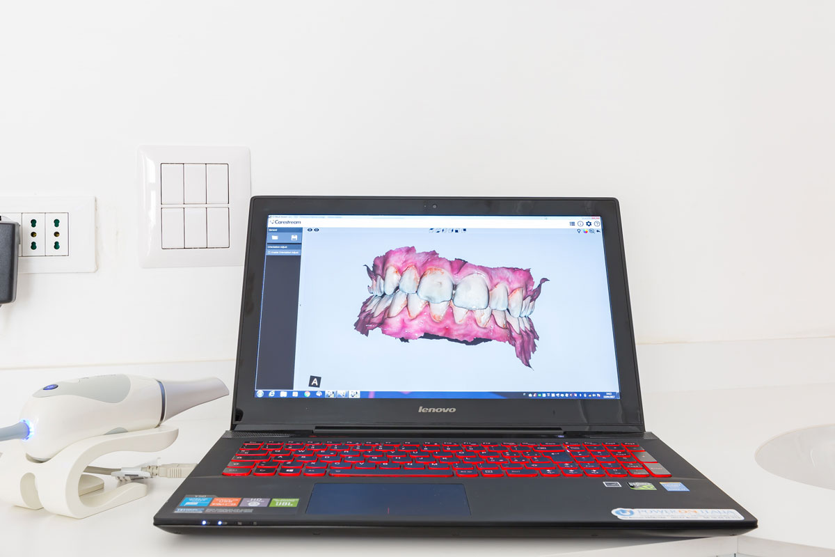
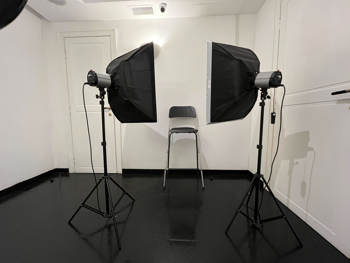
Photo Studio
The photography studio allows all team specialists to document various phases of diagnosis and treatment optimally. Through digital photography, we can capture initial information of our clinical cases and, using dedicated software, study rehabilitation possibilities, anticipating aesthetic results for our patients. The photography studio is an essential communication tool between the clinician and the patient, guiding them through image analysis towards a more informed treatment choice. Photographic analysis places the patient at the center of the therapeutic decision-making process.
